Malignant Melanoma is a skin cancer accounting for around 3% of skin cancer in the UK. It is a malignant tumour arising from the melanocytes – the cells that produce brown pigment and moles. When melanocytes become malignant they form melanoma, but a pre-cancerous variant called lentigo maligna exists. In the early stages, melanoma can be slow-growing, in later stages it has the potential to spread (or metastasise) to other areas of the body.
Alternative names: Melanoma, skin cancer, cancerous mole.
REQUEST A CALL BACK
Arrange a consultation with one of our expert dermatologists today.
WHAT DOES MALIGNANT MELANOMA LOOK LIKE?
Melanomas tend to resemble a mole that is undergoing changes in size, shape or colour. They may become lumpy or ‘nodular’ and even ulcerate with bleeding. Sometimes they occur in a pre-existing mole, but around half the time they arise in an area or normal skin.
Pre-melanoma (lentigo maligna) and the earliest phase of melanoma (in-situ melanoma) are almost always cured with surgery. For melanoma beyond these early stages, it is the depth into the skin that the melanoma reaches that tells us how the disease might behave. This depth is termed the ’Breslow thickness’. The Breslow thickness will determine whether you require a simple excision or further more complex surgery including lymph gland biopsy.
The degree of spread a melanoma has undergone will be classified by stages:
Stage I: Localised to the original site only
Stage II: Localised to the skin only – the original site plus local skin spread
Stage III: Spread to the nearby lymph glands
Stage IV: Spread to the internal organs of the body
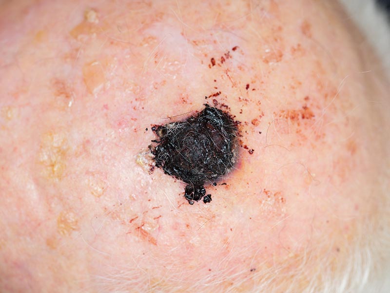
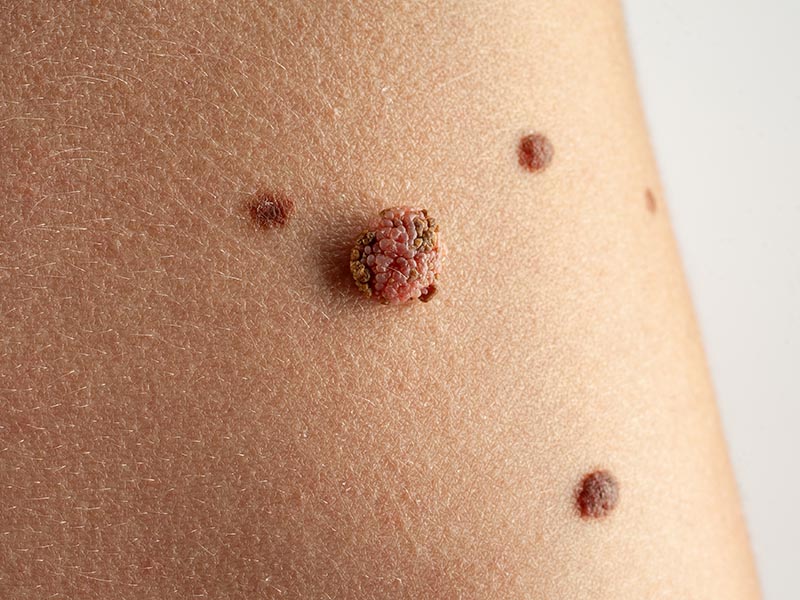
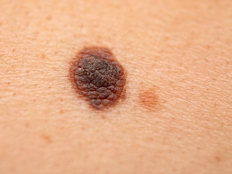
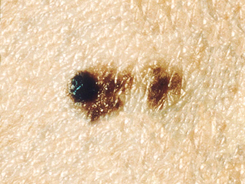
SIGNS OF MALIGNANT MELANOMA
It is a good idea to check your moles once a month, especially if you have lots of moles or freckles (particularly if some are large), have fair hair or skin, use sunbeds or have a family history of skin cancer. When checking your moles, it is a good idea to use both a full length and a hand held mirror so you are able to check your body all over. Stand in a well lit room and ask a family member or your partner to help you check the hard to reach areas. Don’t forget to check less obvious places such as your scalp, the soles of your feet and in between your fingers and toes. It is a good idea to take photos of your moles so you can keep track of any changes. If you do notice any changes, consult a dermatologist.
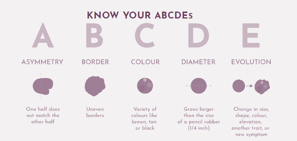
Moles can look very different to each other which can make it hard to identify moles which may be worrying, the key thing you are looking for is if your mole is changing. When checking your body for moles, you are looking for any changes to the size, colour of shape. You are also looking for itching, bleeding or crusting of moles which are signs you need to book an appointment to get your moles checked by a consultant dermatologist. When checking for moles, know your ABCDEs and get your moles professional checked if:
A: Asymmetry – the mole looks unusual, asymmetrical or irregular
B: Border – the border becomes blurred, ill-defined or irregular
C: Colour – more than 2 colours appear in the mole including brown, black or light areas giving a mottled appearance. A very dark or black appearance can occur
D: Diameter – if a mole is enlarging it should be reviewed
HOW CAN MALIGNANT MELANOMA BE TREATED?
All melanomas are treated by excision. The tumour is then assessed under the microscope and the next step will depend on the Breslow thickness. With the diagnosis confirmed, a further operation will be required to excise the scar (wide local excision). This extent of this wide local will depend on the Breslow thickness (as does the likelihood of spread or recurrence). If this is greater than 1 mm, a biopsy of the lymph glands will also be required. Should there be melanoma present in the lymph glands, further surgery will be required.
Remember that the earlier your melanoma is diagnosed
and treated, the less the cosmetic disfigurement and
the less chance of recurrence or spread.
For more information on malignant melanoma, please see the British Association of Dermatologists website melanoma advice leaflets for stage 1, stage 2, stage 3, and stage 4 melanoma.
FREQUENTLY ASKED QUESTIONS
What are the stages of malignant melanoma?
Pre-melanoma (lentigo maligna) and the earliest phase of melanoma (in-situ melanoma) are almost always cured with surgery. For melanoma beyond these early stages, it is the depth into the skin that the melanoma reaches that tells us how the disease might behave. This depth is termed the ’Breslow thickness’. The Breslow thickness will determine whether you require a simple excision or furthermore complex surgery including lymph gland biopsy.
The degree of spread a melanoma has undergone will be classified by stages:
Stage I: Localised to the original site only
Stage II: Localised to the skin only – the original site plus local skin spread
Stage III: Spread to the nearby lymph glands
Stage IV: Spread to the internal organs of the body
What does malignant melanoma look like?
Melanomas tend to resemble a mole that is undergoing changes in size, shape or colour. They may become lumpy or ‘nodular’ and even ulcerate with bleeding. Sometimes they occur in a pre-existing mole, but around half the time they arise in an area or normal skin.
Am I more likely to get malignant melanoma if i've had it before?
If you have already had melanoma, there is a chance it may return again, particularly if the cancer was more advanced or widespread. Your doctors will discuss this with you and will regularly monitor you for signs it has returned.
What should I be looking out for when I check my moles?
A: Asymmetry – the mole looks unusual, asymmetrical or irregular
B: Border – the border becomes blurred, ill-defined or irregular
C: Colour – more than 2 colours appear in the mole including brown, black or light areas giving a mottled appearance. A very dark or black appearance can occur
D: Diameter – if a mole is getting bigger it should be reviewed.
E: Evolving – if a mole changes it should be reviewed
WHY CHOOSE THE HARLEY STREET DERMATOLGY CLINIC?
Having the right dermatologist is important especially when you have a chronic skin condition that will require ongoing treatment. We want you to feel confident that we’re providing you with the best possible care. We also want you to feel as comfortable as possible with your dermatologist.
The Harley Street Dermatology Clinic specialises in conditions affecting the skin, hair and nails. Our goal is to provide all the care that you need when you’re experiencing these kinds of problems. We want to make it easy for you to access the best quality treatment and support in London.
The clinic is conveniently located in Central London, so it’s easy to visit us if you need to see a dermatologist. You will find yourself in a very comfortable and welcoming environment. We have created a relaxing space where you will receive the highest quality of care. We are regulated by the Care Quality Commission, are part of the British Association of Dermatologists and are top rated by patients of Doctify so you can be sure of safe and effective treatment with us.
CONTACT US
Finding Us
The Harley Street Dermatology Clinic
35 Devonshire Place
London
W1G 6JP
The clinic can be accessed by public transport, on foot or by car. There is paid on street parking around the Harley Street district. The nearest tube stations are Regent’s Park and Baker Street, and Marylebone train station is a 15 minute walk away.
Contact Details
Opening Hours
While appointments can be made available outside usual hours in special circumstances, our core hours are:
Monday: 8am - 6pm
Tuesday: 8am - 6pm
Wednesday: 8am - 6pm
Thursday: 8am - 6pm
Friday: 8am - 6pm
Saturday : 10am - 2pm
Sunday: Closed
REQUEST A CALL BACK
Please fill in this form and one of our team will give you a call back to arrange a consultation with one of our expert dermatologists.
